PLD3
| PLD3 | |||||||||||||||||||||||||||||||||||||||||||||||||||
|---|---|---|---|---|---|---|---|---|---|---|---|---|---|---|---|---|---|---|---|---|---|---|---|---|---|---|---|---|---|---|---|---|---|---|---|---|---|---|---|---|---|---|---|---|---|---|---|---|---|---|---|
| Identifiers | |||||||||||||||||||||||||||||||||||||||||||||||||||
| Aliases | PLD3, AD19, HU-K4, HUK4, phospholipase D family member 3, SCA46 | ||||||||||||||||||||||||||||||||||||||||||||||||||
| External IDs | OMIM: 615698; MGI: 1333782; HomoloGene: 7893; GeneCards: PLD3; OMA:PLD3 - orthologs | ||||||||||||||||||||||||||||||||||||||||||||||||||
| |||||||||||||||||||||||||||||||||||||||||||||||||||
| |||||||||||||||||||||||||||||||||||||||||||||||||||
| |||||||||||||||||||||||||||||||||||||||||||||||||||
| |||||||||||||||||||||||||||||||||||||||||||||||||||
| |||||||||||||||||||||||||||||||||||||||||||||||||||
| Wikidata | |||||||||||||||||||||||||||||||||||||||||||||||||||
| |||||||||||||||||||||||||||||||||||||||||||||||||||
Phospholipase D3, also known as PLD3, is a protein that in humans is encoded by the PLD3 gene.[5][6] PLD3 belongs to the phospholipase D superfamily because it contains the two HKD motifs common to members of the phospholipase D family, however, it has no known catalytic function similar to PLD1 or PLD2. PLD3 serves as a ssDNA 5' exonuclease in antigen presenting cells.[7] PLD3 is highly expressed in the brain in both humans and mice, and is mainly localized in the endoplasmic reticulum (ER) and the lysosome.
PLD3 may play a role in regulating the lysosomal system, myogenesis, late-stage neurogenesis, inhibiting insulin signal transduction, and amyloid precursor protein (APP) processing. The involvement in PLD3 in the lysosomal system and in APP processing and the loss-of-function mutations in PLD3 are thought to be linked to late-onset Alzheimer's disease (LOAD).[8][9] However, there are also studies that challenge the association between PLD3 and Alzheimer's disease (AD).[10][11][12][13][14]
How APP processing is affected by PLD3 during AD still remains unclear, and its role in the pathogenesis of AD is ambiguous.[14][15] PLD3 may contribute to the onset of AD by a mechanism other than by influencing APP metabolism, with one proposed mechanism suggesting that PLD3 contributes to the onset of AD by impairing the endosomal-lysosomal system.[14] In 2017, PLD3 was shown to have an association with another neurodegenerative disease, spinocerebellar ataxia.[16]
Genetics
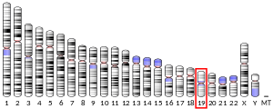
[edit]PLD3 was first characterized as a human homolog of the HindIII K4L protein in the vaccinia virus, having a DNA sequence 48.1% similar to the viral gene.[17] The PLD3 gene in humans is located at chromosome 19q13.2, with a sequence comprising at least 15 exons and is alternatively spliced at the low GC 5' UTR into 25 predicted transcripts.[18][19] Translation of the 490 amino acid-long PLD3 protein is initiated around exons 5 to 7, and ends at the stop codon in exon 15.[18]
Structure
[edit]PLD3 is a 490 amino acid-long type 2 transmembrane protein, unlike PLD1 and PLD2 which do not contain a transmembrane protein domain in their protein structure.[18]
The cytosolic N-terminal of the protein faces towards the cytoplasm of the cell, and lacks consensus sites for N-glycosylation.[18] The N-terminus is also predicted to contain a transmembrane domain.[20]
The bulk of the protein is located in the ER lumen, containing the C-terminal domain.[21] The C-terminal domain contains seven glycosylation sites along with a prenylation motif and two HXKXXXXD/E (HKD) motifs.[18] In PLD1 and PLD2, this is the catalytic domain or active site of the protein, which is why PLD3 was assigned to the phospholipase D superfamily.[18] However, PLD3 has no known catalytic activity and aside from presence of the HKD motifs, PLD3 has no structural commonalities with PLD1 or PLD2.[18]
Tissue and subcellular distribution
[edit]Expression of PLD3 in tissues differs with the transcript size of its mRNA.[18] The longer 2200 base pair transcript is ubiquitously expressed in the body, exhibiting higher expression levels in the heart, skeletal muscle, and the brain.[18] Meanwhile, the shorter 1700 base pair transcript is found in abundance in the brain, but at low expression in non-nervous tissue.[18][22] PLD3 expression is especially pronounced in mature neurons in the mammalian forebrain.[22] High expression of PLD3 is specifically seen in the hippocampus and the frontal, temporal, and occipital lobes in the cerebral cortex.[8][22] The PLD3 gene is also found with high expression in the cerebellum.[16]
Subcellular localization of PLD3 is thought to primarily be in the endoplasmic reticulum (ER), as it has been shown to co-localize with protein disulfide-isomerase, a protein known to be a marker for the ER.[18] PLD3 may also be localized in lysosomes, co-localizing with lysosomal markers LAMP1 and LAMP2 in lysosomes in separate studies.[14][23] PLD3 was identified as a protein in insulin secretory granules derived from pancreatic beta cells.[24]
Function
[edit]PLD3 is a member of the phospholipase D protein family, however, unlike phospholipase PLD1 and PLD2,[18] it serves as a 5' exonuclease that specifically degrade ssDNA in the endolysosome, which is similar to the function of PLD4. Both PLD3 and PLD4 are essential for the clearance of nucleic acid product in antigen presenting cells. Deletion of PLD3 and PLD4 leads to accumulation of ssDNA and RNA in the endosome, which activates various nucleic acid sensors including TLR9, TLR7 and cGAS-STING and triggers inflammation and elevated secretion of cytokines.[7][25] It is shown that mitochondrial DNA (mtDNA) is the major physiological substrate for PLD3 to degrade. [26]
PLD3 may play some role in influencing protein processing through the lysosome as well as a regulatory role in lysosomal morphology.[14] Some studies suggest that PLD3 is involved in amyloid precursor protein (APP) processing and regulating amyloid beta (Aβ) levels.[8] Overexpression of wildtype PLD3 is linked to a decrease in intracellular APP and extracellular Aβ isoforms Aβ40 and Aβ42, while a knockdown of PLD3 is linked to an increase in extracellular Aβ40 and Aβ42.[8] PLD3 was implied to be involved in sensing oxidative stress, such that suppressing the PLD3 gene with short hairpin RNA increased the viability of cells exposed to oxidative stress.[27]
Increased PLD3 expression was shown to increase myotube formation in differentiated mouse myoblasts in vitro, and ER stress which also increases myotube formation was also shown to increase PLD3 expression.[20] Decreasing PLD3 expression meanwhile decreases myotube formation.[20] These findings suggest a possible role of PLD3 in myogenesis, although its exact mechanism of action remains unknown.[20] Overexpression of PLD3 in mouse myoblasts in vitro may inhibit Akt phosphorylation and block signal transduction during insulin signalling.[28] PLD3 may be involved in the later stages of neurogenesis, contributing to processes associated with neurotransmission, target cell innervation, and neuronal survival.[22]
Elevated expression of PLD3 was found to be one of the consistent factors that contribute to the self-renewal activity of hematopoietic stem cell populations, suggesting a possible role of PLD3 in the mechanism behind the maintenance of durable, long-term self-renewing cell populations.[29]
Interactions
[edit]The human progranulin protein (PGRN), encoded by the human granulin gene (GRN), is co-expressed with and interacts with PLD3 accumulated on neuritic plaques in AD brains.[30] PLD3 may interact with APP and amyloid beta, as some studies indicate that PLD3 is involved with APP processing and regulating Aβ levels.[8] PLD3 may also interact with Akt and insulin in myoblasts in vitro.[28]
Clinical significance
[edit]Alzheimer's disease
[edit]Mutations in PLD3 have been studied for their potential role in the pathogenesis of late-onset Alzheimer's disease (LOAD).[8]
In 2013, Cruchaga et al. found that a particular rare coding variant or missense mutation in PLD3 (Val232Met) doubled the risk for Alzheimer's disease among cases and controls of European and African-American descent.[8] PLD3 mRNA and protein expression was reduced in AD brains compared with non-AD brains in regions that PLD3 is normally found with high expression, and another study also found that PLD3 accumulates on neuritic plaques in AD brains.[8][30] A common PLD3 single nucleotide polymorphism (SNP) was also found to have an association with Aβ pathology among normal, healthy individuals, suggesting that common PLD3 variants may also be involved in the pathogenesis of AD.[31] A meta-analysis conducted in 2015 concluded that the Val232Met PLD3 variant has a modest effect on increasing AD risk.[9]
However, the findings from Cruchaga et al. could not be replicated in follow-up studies on the role of PLD3 in both familial and non-familial, sporadic Alzheimer's disease in Western population samples.[10][11][12] The Val232Met PLD3 mutant was also not identified in a sample of AD patients and healthy control subjects from mainland China, suggesting that this particular PLD3 mutant may not significantly affect AD risk in the mainland Chinese population.[13] A study showed that while there is an excess of PLD3 variants in LOAD, none of the variants described by Cruchaga et al. drive the association between PLD3 and LOAD in a European cohort, including the Val232Met variant.[32] This study along with an additional study also demonstrated that these rare coding variants of PLD3 were not observed in early-onset AD (EOAD) in a European cohort, suggesting that PLD3 may not have a role in EOAD.[32][33]
The underlying mechanisms on how mutations in PLD3 affects APP processing in AD remains unclear.[15] Results from the study by Cruchaga et al. indicated that PLD3 loss-of-function increases risk for Alzheimer's disease by affecting APP processing.[8] The involvement of PLD3 in APP processing was challenged in a recent study which showed that a PLD3 loss-of-function does not significantly affect the burden of amyloid plaques on AD development in mice.[14] PLD3 loss-of-function in this study did, however, change the morphology of the lysosomal system in neurons, indicating that PLD3 loss-of-function may still be involved in the pathophysiology of AD through some other mechanism such as by contributing to the impairment of the endosomal-lysosomal system that occurs during AD.[34][35]
Spinocerebellar ataxia
[edit]In 2017, the PLD3 gene was identified as one of the novel genes linked to spinocerebellar ataxia, another neurodegenerative genetic disease.[16]
References
[edit]- ^ a b c GRCh38: Ensembl release 89: ENSG00000105223 – Ensembl, May 2017
- ^ a b c GRCm38: Ensembl release 89: ENSMUSG00000003363 – Ensembl, May 2017
- ^ "Human PubMed Reference:". National Center for Biotechnology Information, U.S. National Library of Medicine.
- ^ "Mouse PubMed Reference:". National Center for Biotechnology Information, U.S. National Library of Medicine.
- ^ "Entrez Gene: Phospholipase D family, member 3".
- ^ Universal protein resource accession number Q8IV08 for "PLD3 - Phospholipase D3 - Homo sapiens" at UniProt.
- ^ a b Gavin AL, Huang D, Huber C, et al. (September 2018). "PLD3 and PLD4 are single-stranded acid exonucleases that regulate endosomal nucleic-acid sensing". Nature Immunology. 19 (9): 942–953. doi:10.1038/s41590-018-0179-y. PMC 6105523. PMID 30111894.
- ^ a b c d e f g h i Cruchaga C, Karch CM, Jin SC, et al. (January 2014). "Rare coding variants in the phospholipase D3 gene confer risk for Alzheimer's disease". Nature. 505 (7484): 550–554. Bibcode:2014Natur.505..550.. doi:10.1038/nature12825. PMC 4050701. PMID 24336208.
- ^ a b Zhang DF, Fan Y, Wang D, et al. (August 2016). "PLD3 in Alzheimer's Disease: a Modest Effect as Revealed by Updated Association and Expression Analyses". Molecular Neurobiology. 53 (6): 4034–4045. doi:10.1007/s12035-015-9353-5. PMID 26189833. S2CID 15660705.
- ^ a b Heilmann S, Drichel D, Clarimon J, et al. (April 2015). "PLD3 in non-familial Alzheimer's disease". Nature. 520 (7545): E3-5. Bibcode:2015Natur.520E...3H. doi:10.1038/nature14039. PMID 25832411. S2CID 205241800.
- ^ a b Hooli BV, Lill CM, Mullin K, et al. (April 2015). "PLD3 gene variants and Alzheimer's disease". Nature. 520 (7545): E7-8. Bibcode:2015Natur.520E...7H. doi:10.1038/nature14040. PMID 25832413. S2CID 4388686.
- ^ a b Lambert JC, Grenier-Boley B, Bellenguez C, et al. (April 2015). "PLD3 and sporadic Alzheimer's disease risk". Nature. 520 (7545): E1. Bibcode:2015Natur.520E...1L. doi:10.1038/nature14036. PMID 25832408. S2CID 52869488.
- ^ a b Jiao B, Liu X, Tang B, et al. (October 2014). "Investigation of TREM2, PLD3, and UNC5C variants in patients with Alzheimer's disease from mainland China". Neurobiology of Aging. 35 (10): 2422.e9–2422.e11. doi:10.1016/j.neurobiolaging.2014.04.025. PMID 24866402. S2CID 140208846.
- ^ a b c d e f Fazzari P, Horre K, Arranz AM, et al. (January 2017). "PLD3 gene and processing of APP" (PDF). Nature. 541 (7638): E1–E2. Bibcode:2017Natur.541E...1F. doi:10.1038/nature21030. PMID 28128235. S2CID 1131827.
- ^ a b Wang J, Yu JT, Tan L (April 2015). "PLD3 in Alzheimer's disease". Molecular Neurobiology. 51 (2): 480–6. doi:10.1007/s12035-014-8779-5. PMID 24935720. S2CID 15700225.
- ^ a b c Nibbeling EA, Duarri A, Verschuuren-Bemelmans CC, et al. (November 2017). "Exome sequencing and network analysis identifies shared mechanisms underlying spinocerebellar ataxia". Brain. 140 (11): 2860–2878. doi:10.1093/brain/awx251. PMID 29053796.
- ^ Cao JX, Koop BF, Upton C (April 1997). "A human homolog of the vaccinia virus HindIII K4L gene is a member of the phospholipase D superfamily". Virus Research. 48 (1): 11–8. doi:10.1016/s0168-1702(96)01422-0. PMID 9140189.
- ^ a b c d e f g h i j k l Munck A, Böhm C, Seibel NM, et al. (April 2005). "Hu-K4 is a ubiquitously expressed type 2 transmembrane protein associated with the endoplasmic reticulum". The FEBS Journal. 272 (7): 1718–26. doi:10.1111/j.1742-4658.2005.04601.x. PMID 15794758. S2CID 22592569.
- ^ Karch CM, Goate AM (January 2015). "Alzheimer's disease risk genes and mechanisms of disease pathogenesis". Biological Psychiatry. 77 (1): 43–51. doi:10.1016/j.biopsych.2014.05.006. PMC 4234692. PMID 24951455.
- ^ a b c d Osisami M, Ali W, Frohman MA (2012-03-12). "A role for phospholipase D3 in myotube formation". PLOS ONE. 7 (3): e33341. Bibcode:2012PLoSO...733341O. doi:10.1371/journal.pone.0033341. PMC 3299777. PMID 22428023.
- ^ Frohman MA (March 2015). "The phospholipase D superfamily as therapeutic targets". Trends in Pharmacological Sciences. 36 (3): 137–44. doi:10.1016/j.tips.2015.01.001. PMC 4355084. PMID 25661257.
- ^ a b c d Pedersen KM, Finsen B, Celis JE, et al. (November 1998). "Expression of a novel murine phospholipase D homolog coincides with late neuronal development in the forebrain". The Journal of Biological Chemistry. 273 (47): 31494–504. doi:10.1074/jbc.273.47.31494. PMID 9813063.
- ^ Palmieri M, Impey S, Kang H, et al. (October 2011). "Characterization of the CLEAR network reveals an integrated control of cellular clearance pathways". Human Molecular Genetics. 20 (19): 3852–66. doi:10.1093/hmg/ddr306. PMID 21752829.
- ^ Brunner Y, Couté Y, Iezzi M, et al. (June 2007). "Proteomics analysis of insulin secretory granules". Molecular & Cellular Proteomics. 6 (6): 1007–17. doi:10.1074/mcp.M600443-MCP200. PMID 17317658.
- ^ Gavin AL, Huang D, Blane TR, et al. (October 2021). "Cleavage of DNA and RNA by PLD3 and PLD4 limits autoinflammatory triggering by multiple sensors". Nature Communications. 12 (1): 5874. Bibcode:2021NatCo..12.5874G. doi:10.1038/s41467-021-26150-w. PMC 8497607. PMID 34620855.
- ^ Van Acker ZP, Perdok A, Hellemans R, et al. (May 2023). "Phospholipase D3 degrades mitochondrial DNA to regulate nucleotide signaling and APP metabolism". Nature Communications. 14 (1): 2847. Bibcode:2023NatCo..14.2847V. doi:10.1038/s41467-023-38501-w. PMC 10209153. PMID 37225734.
- ^ Nagaoka-Yasuda R, Matsuo N, Perkins B, et al. (September 2007). "An RNAi-based genetic screen for oxidative stress resistance reveals retinol saturase as a mediator of stress resistance". Free Radical Biology & Medicine. 43 (5): 781–8. doi:10.1016/j.freeradbiomed.2007.05.008. PMID 17664141.
- ^ a b Zhang J, Chen S, Zhang S, et al. (October 2009). "[Over-expression of phospholipase D3 inhibits Akt phosphorylation in C2C12 myoblasts]". Sheng Wu Gong Cheng Xue Bao = Chinese Journal of Biotechnology. 25 (10): 1524–31. PMID 20112697.
- ^ Kent DG, Copley MR, Benz C, et al. (June 2009). "Prospective isolation and molecular characterization of hematopoietic stem cells with durable self-renewal potential". Blood. 113 (25): 6342–50. doi:10.1182/blood-2008-12-192054. PMID 19377048.
- ^ a b Satoh J, Kino Y, Yamamoto Y, et al. (2014-11-02). "PLD3 is accumulated on neuritic plaques in Alzheimer's disease brains". Alzheimer's Research & Therapy. 6 (9): 70. doi:10.1186/s13195-014-0070-5. PMC 4255636. PMID 25478031.
- ^ Wang C, Tan L, Wang HF, et al. (2015). "Common Variants in PLD3 and Correlation to Amyloid-Related Phenotypes in Alzheimer's Disease". Journal of Alzheimer's Disease. 46 (2): 491–5. doi:10.3233/JAD-150110. PMC 6312181. PMID 26402410.
- ^ a b Schulte EC, Kurz A, Alexopoulos P, et al. (2015-01-09). "Excess of rare coding variants in PLD3 in late- but not early-onset Alzheimer's disease". Human Genome Variation. 2: 14028. doi:10.1038/hgv.2014.28. PMC 4785568. PMID 27081517.
- ^ Cacace R, Van den Bossche T, Engelborghs S, et al. (December 2015). "Rare Variants in PLD3 Do Not Affect Risk for Early-Onset Alzheimer Disease in a European Consortium Cohort". Human Mutation. 36 (12): 1226–35. doi:10.1002/humu.22908. PMC 5057316. PMID 26411346.
- ^ Nixon RA (August 2013). "The role of autophagy in neurodegenerative disease". Nature Medicine. 19 (8): 983–997. doi:10.1038/nm.3232. PMID 23921753. S2CID 9152845.
- ^ Cataldo AM, Hamilton DJ, Barnett JL, et al. (January 1996). "Properties of the endosomal-lysosomal system in the human central nervous system: disturbances mark most neurons in populations at risk to degenerate in Alzheimer's disease". The Journal of Neuroscience. 16 (1): 186–199. doi:10.1523/JNEUROSCI.16-01-00186.1996. PMC 6578706. PMID 8613784.
Further reading
[edit]- Idkowiak-Baldys J, Baldys A, Raymond JR, et al. (August 2009). "Sustained receptor stimulation leads to sequestration of recycling endosomes in a classical protein kinase C- and phospholipase D-dependent manner". The Journal of Biological Chemistry. 284 (33): 22322–31. doi:10.1074/jbc.M109.026765. PMC 2755955. PMID 19525236.
- Miyamoto-Sato E, Fujimori S, Ishizaka M, et al. (February 2010). "A comprehensive resource of interacting protein regions for refining human transcription factor networks". PLOS ONE. 5 (2): e9289. Bibcode:2010PLoSO...5.9289M. doi:10.1371/journal.pone.0009289. PMC 2827538. PMID 20195357.
- Walker LC, Waddell N, Ten Haaf A, et al. (November 2008). "Use of expression data and the CGEMS genome-wide breast cancer association study to identify genes that may modify risk in BRCA1/2 mutation carriers". Breast Cancer Research and Treatment. 112 (2): 229–36. doi:10.1007/s10549-007-9848-5. PMID 18095154. S2CID 795870.
- Pridgeon JW, Webber EA, Sha D, et al. (January 2009). "Proteomic analysis reveals Hrs ubiquitin-interacting motif-mediated ubiquitin signaling in multiple cellular processes". The FEBS Journal. 276 (1): 118–31. doi:10.1111/j.1742-4658.2008.06760.x. PMC 2647816. PMID 19019082.
- Russo P, Venezia A, Lauria F, et al. (April 2006). "HindIII(+/-) polymorphism of the Y chromosome, blood pressure, and serum lipids: no evidence of association in three white populations". American Journal of Hypertension. 19 (4): 331–8. doi:10.1016/j.amjhyper.2005.10.003. PMID 16580565.
- Cheng J, Kapranov P, Drenkow J, et al. (May 2005). "Transcriptional maps of 10 human chromosomes at 5-nucleotide resolution". Science. 308 (5725): 1149–54. Bibcode:2005Sci...308.1149C. doi:10.1126/science.1108625. PMID 15790807. S2CID 13047538.
This article incorporates text from the United States National Library of Medicine, which is in the public domain.


