Jump to content

File:Balo sclerosis.JPG

Page contents not supported in other languages.
This is a file from the Wikimedia Commons
From Wikipedia, the free encyclopedia

Original file (1,494 × 1,277 pixels, file size: 303 KB, MIME type: image/jpeg)

Description

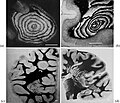
Typical aspects of Baló's concentric sclerosis. (a) Original case of Baló; several anastomoses are located in the lower half of the lesion (from Baló (1928) Arch Neurol Psychiatr 19:242–264). (b) Lesion centered by a veinule showing ring fragmentation in a constrained area (from Hallervorden et al. 1933). (c) Lesion reproduced from Castaigne et al. (1984) Rev Neurol 140:479–487. (d) Progress of the pathologic process from a center located in a constrained area, showing formation of bands (from Behr (1950) Dtsch Z Nervenheilk 164:480–489). Loyez staining (myelin in black, destroyed areas in white); scale bars: 1 cm.

doi:10.1371/journal.pone.0000150.g001
Date 10 March 2008 (upload date)
Source The Origins of Concentric Demyelination: Self-Organization in the Human Brain. Roman H. Khonsari and Vincent Calvez PLoS ONE. 2007; 2(1): e150. [1]
Author Roman H. Khonsari, Vincent Calvez
w:en:Creative Commons
attribution
This file is licensed under the Creative Commons Attribution 1.0 Generic license.
You are free:
  • to share – to copy, distribute and transmit the work
  • to remix – to adapt the work
Under the following conditions:
  • attribution – You must give appropriate credit, provide a link to the license, and indicate if changes were made. You may do so in any reasonable manner, but not in any way that suggests the licensor endorses you or your use.

Captions

Add a one-line explanation of what this file represents

Items portrayed in this file

depicts

10 March 2008

image/jpeg

15e2f79456002b9a2d69ed5eb819a0695ecaf7b6

310,485 byte

1,277 pixel

1,494 pixel

File history

Click on a date/time to view the file as it appeared at that time.

Date/TimeThumbnailDimensionsUserComment
current17:24, 10 March 2008Thumbnail for version as of 17:24, 10 March 20081,494 × 1,277 (303 KB)Filip em{{Information |Description=Typical aspects of Baló's concentric sclerosis. (a) Original case of Baló; several anastomoses are located in the lower half of the lesion (from Baló (1928) Arch Neurol Psychiatr 19:242–264). (b) Lesion centered by a veinul

The following page uses this file:

Global file usage

Metadata